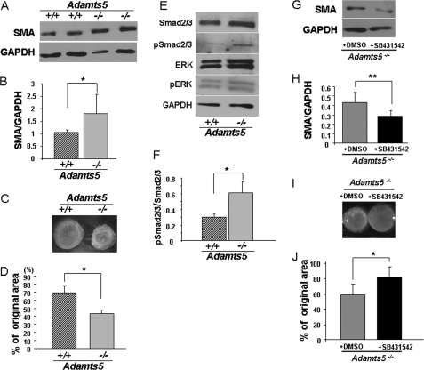
FIGURE 3.
Adamts5−/− dermal fibroblasts show features of myofibroblasts. A, Western blot for SMA shows increased levels in Adamts5−/− dermal fibroblasts (−/−) compared with WT (+/+) (representative of three biological replicates and five technical replicates). B, results of densitometric analysis of SMA Western blots show a statistically significant increase in Adamts5−/− dermal fibroblasts (*, p < 0.01). C, a collagen gel contraction assay (representative example) shows the greater contractility of Adamts5−/−dermal fibroblasts. D, quantification of gel contraction assay shows a statistically significant difference (*, p < 0.01) in contractility between Adamts5−/− and WT dermal fibroblasts (n = 3 biological replicates, 4 technical replicates, and triplicate wells per experiment). E, Western blot analyses with the indicated antibodies show increased pSmad levels but no change in pERK in Adamts5−/− dermal fibroblasts (representative of n = 3). F, quantification by densitometric analysis of pSmad2/3 to total Smad2/3 shows a statistically significant difference between Adamts5−/− and WT dermal fibroblasts (*, p < 0.01) (n = 3 biological replicates). Error bars indicate S.D. G, Adamts5−/− cells were treated with SB431542, which led to decreased expression of SMA (representative of n = 3). H, quantification of SMA showed statistically significant reduction by 4MU-treated cells (**, p < 0.05). I, the collagen gel contraction assay was done using Adamts5−/− cells treated with SB431542 or with DMSO (vehicle for SB431542 delivery) as a control. J, a statistically significant reduction of contractility was observed in SB431542-treated Adamts5−/− cells (*, p < 0.01, n = 3). Error bars indicate S.D.

