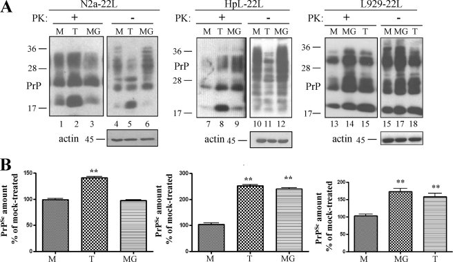
FIGURE 5.
ER stress and proteasomal dysfunction enhance accumulation of PrPSc in prion-infected cells. A, the amount of PrPSc in N2a-22l, HpL-22L, and L929-22L cells treated with tunicamycin (T) or MG132 (MG) was compared with that in mock treated (M) cells. Immunoblots show total PrP- (−PK) and PrPSc-content (+PK) in post-nuclear lysates probed with mAb 4H11. The lowest panels show actin-loading controls. B, densitometric evaluation of at least two independent experiments performed in triplicate. PrPSc in compound-treated cells was plotted as the percentage of PrPSc measured in mock treated cells (p < 0,005).

