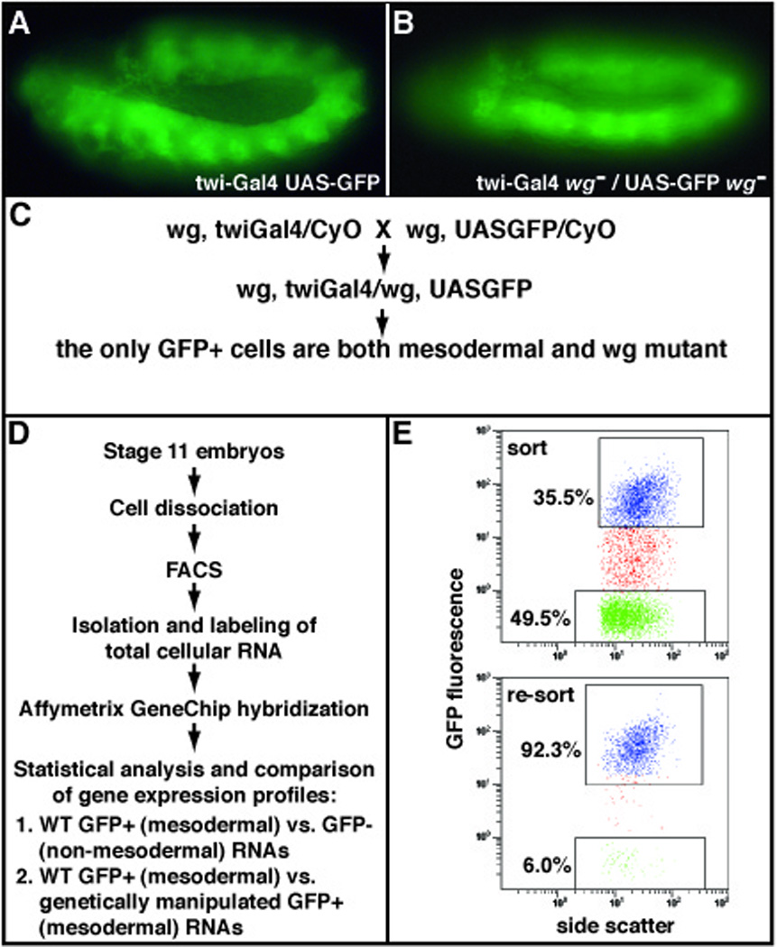
Figure 1. Experimental strategy to obtain gene expression signatures of purified Drosophila embryonic mesodermal cells.

(A) Transgenic, stage 11, Drosophila embryo expressing Gal4 under the control of the twi promoter and GFP under the UAS regulatory sequence (21), resulting in GFP-positive mesodermal cells. (B) Transgenic, stage 11, wingless (wg) mutant embryo with GFP-positive mesodermal cells. (C) Genetic crossing scheme to obtain homozygous mutant GFP-positive mesodermal cells. Strains bearing independently generated recombinant chromosomes having the mutant gene of interest (for example, wg) and either the twiGal4 or UASGFP transgenes are crossed. (D) A representative fluorescence-activated cell sorting (FACS) experiment to obtain total RNA from mesodermally purified cells. (E) Representative FACS scatter plots before (top panel) and after (bottom panel) the separation of GFP-positive and -negative cell populations. Top panel, upper box: GFP-positive sort in blue; top pannel, lower box: GFP-negative sort in green. Bottom panel, upper box: in blue are shown the re-sorted GFP-positive cells to verify that purity obtained from the primary sort was greater than 90%.
