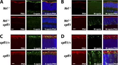
FIGURE 7.
Functional PDE6 is crucial for localization of M-opsin to outer segments. Frozen retinal sections were probed with anti S-opsin (green) and peanut agglutinin (red). S-opsin is present in outer segments irrespective of the functional status of PDE6 (A, C). M -opsin (green) is present in outer segments in Nrl−/− and cpfl1/+ mice (B, D: upper panel) but is mislocalized to synaptic and nuclear layer of retina from Nrl−/− cpfl1 and cpfl1 mice (B, D: lower panel). All retinal sections were from P12 mice. (Scale bar: 10 μm.)

