
FIGURE 2.
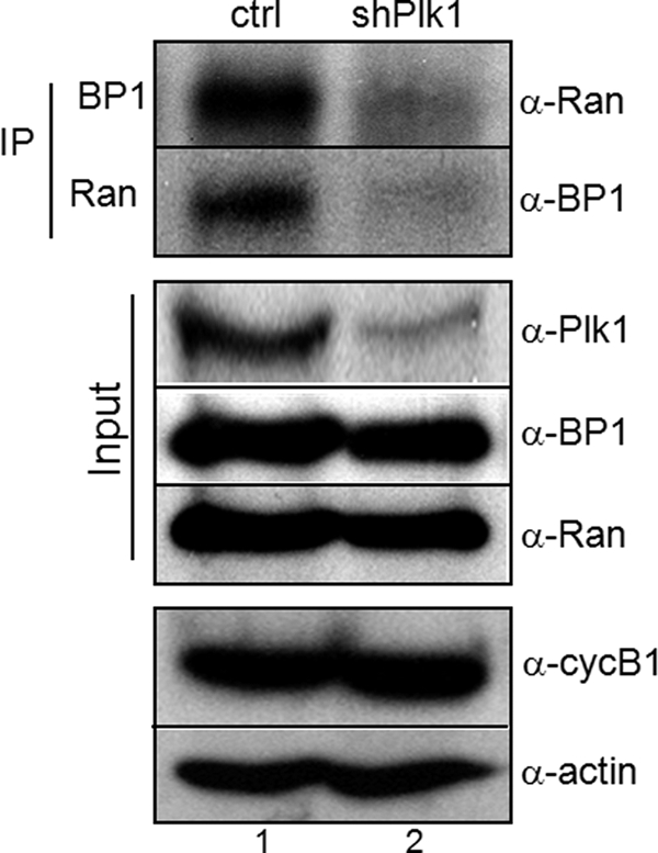
Interaction between Ran and RanBP1 was decreased in depletion of Plk1. In vivo interaction between Ran and RanBP1 was performed by coimmunoprecipitation. Cells were transfected with vector (lane 1, ctrl) or Plk1 shRNA (lane 2, shPlk1). After a 30-h incubation in fresh media, cells were treated with nocodazole for an additional 10 h to arrest in early mitosis. Endogenous Ran and RanBP1 were immunoprecipitated (IP), and bound proteins were detected by immunoblot analysis using appropriate antibodies (top panels, α-Ran or α-BP1). Protein amounts used in this experiment were estimated by actin expression (α-actin).