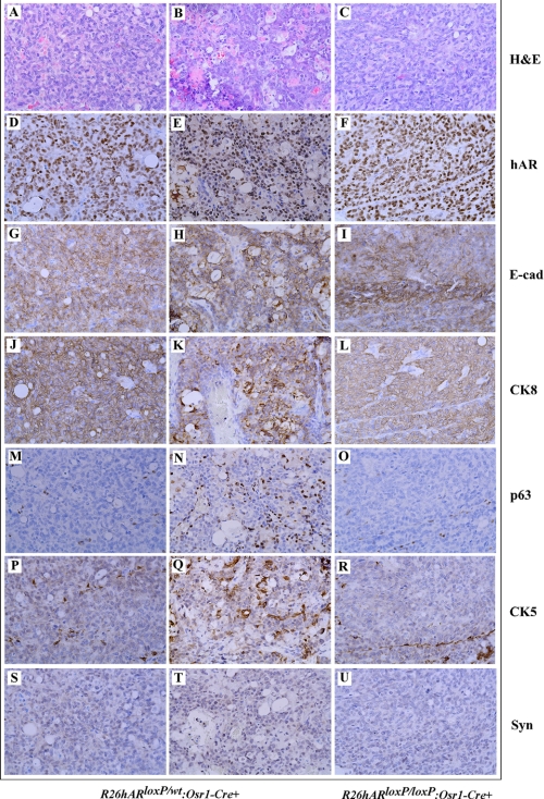
FIGURE 7.
Immunohistochemical analyses of prostate adenocarcinomas in R26hARloxP/wt:Osr1-Cre+ and R26hARloxP/loxP:Osr1-Cre+. Adjacent prostate tissue slides were prepared from prostatic adenocarcinoma regions of R26hARloxP/wt:Osr1-Cre+ and R26hARloxP/loxP:Osr1-Cre+ mice that reported on Fig. 5 and stained with different antibodies as labeled in the figure. A–C, hematoxylin and eosin staining; D–U, immunohistochemistry with different antibodies as labeled in the figure.

