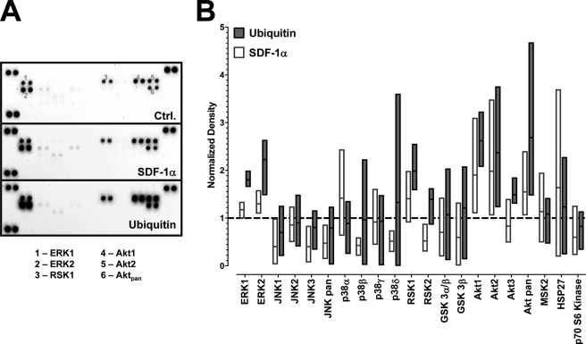
FIGURE 2.
Phosphorylation of mitogen-activated protein kinases after stimulation with SDF-1α and ubiquitin. THP-1 cells were stimulated with 0 or 1 μm ubiquitin or SDF-1α for 10 min at 37 °C. Whole cell lysates were probed for protein kinase phosphorylations utilizing a proteome array. A, proteome array membranes showing the spot densities in untreated (ctrl.), ubiquitin-treated and SDF-1α-treated cells. The numbers on the array membrane correspond to the spot positions for phosphorylated ERK1 (1), ERK2 (2), RSK1 (3), Akt1 (4), Akt2 (5), and Akt pan (6). B, densitometric quantification of the spot densities after treatment as in A, n = 4. Spot densities are given as normalized pixel densities (1 = unstimulated cells, dashed line). The bars (white, SDF-1α treatment; gray, ubiquitin treatment) extend from the minimum to the maximum, the horizontal line shows the mean.

