FIGURE 5.
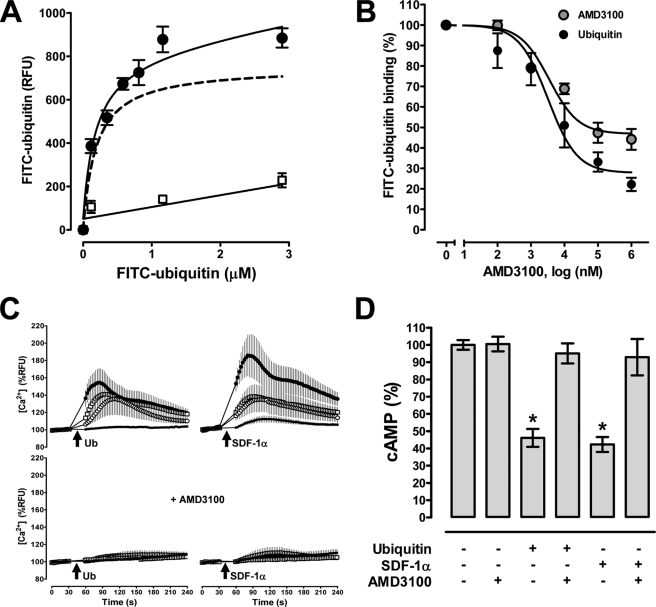
Ubiquitin functions as a CXCR4 agonist in P4.R5 MAGI cells. A, FITC-ubiquitin binding (1 min, 4 °C). ●, FITC-ubiquitin; □, nonspecific binding. Dashed line, specific binding curve (=total FITC-ubiquitin binding − nonspecific binding); n = 6. B, competition binding (1 min, 4 °C) curve for unlabeled ubiquitin (n = 6, ●) and AMD3100 (n = 5, ●) with 1.16 μm FITC-ubiquitin. FITC-ubiquitin binding is expressed as % of the fluorescence signal measured in the absence of unlabeled ubiquitin (=100%). C, top, ubiquitin (left panels) and SDF-1α (right panels) induced Ca2+ flux. Bottom, cells were pretreated with AMD3100 (10 μm); n = 3. Arrows indicate the time point when ubiquitin or SDF-1α was added (●, 1.16 μm; □, 116 nm; ○, 16 nm; ■, 1.6 nm). D, AMD3100 (10 μm) abolishes ubiquitin and SDF-1α (116 nm) induced reduction of cAMP levels in forskolin-stimulated cells; n = 4. Data are expressed as % of untreated cells (=100%). *, p < 0.05 versus untreated cells.

