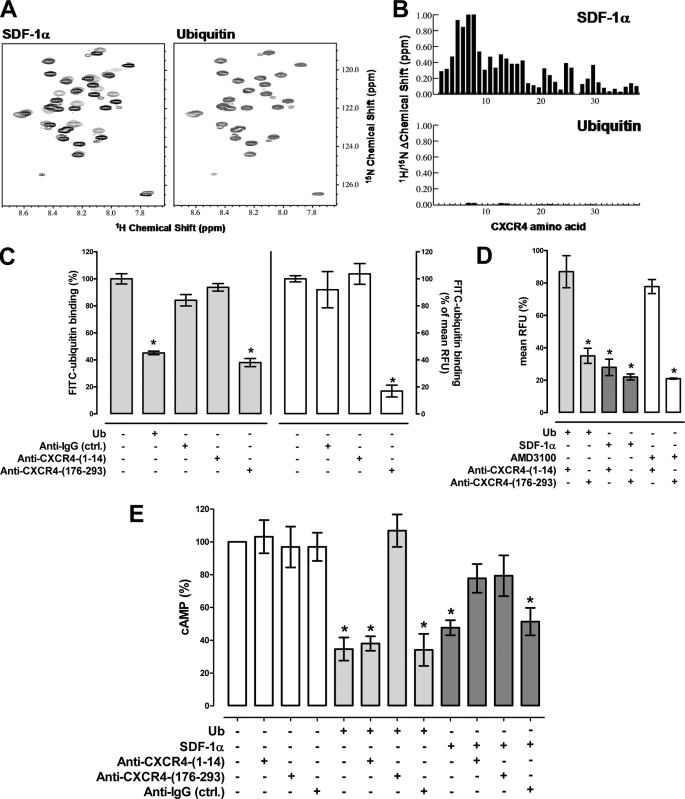
FIGURE 7.
The ubiquitin CXCR4 interaction is independent of the N-terminal receptor domain. A, ubiquitin does not bind CXCR4-(1–38). 15N-1H heteronuclear single quantum coherence of 250 μm [U-15N]CXCR4-(1–38) in the absence (black) and presence (gray) of 325 μm SDF-1α (left) or ubiquitin (right); the chemical shift of CXCR4-(1–38) residues change in the presence of SDF-1α, whereas they are unperturbed by ubiquitin. B, combined 1H/15N shift perturbations of SDF1α (top) and ubiquitin (bottom) plotted as a function of the CXCR4-(1–38) residue. Tyr-7 and Thr-8 were not present at the end of titration with SDF-1α due to line broadening. Shift changes for Pro-27 were not measured because it does not contain an amide proton. C, FITC-ubiquitin binding (1.16 μm) to THP-1 cells after labeling of cells with anti-CXCR4-(1–14), anti-CXCR4-(176–293), or anti-IgG. Gray bars (left y axis), RFU from ubiquitin binding assays. Open bars (right y axis), mean RFU from FACS analyses. Ub, ubiquitin, 30 μm. Data are expressed as % of the RFU after incubation with FITC-ubiquitin alone (=100%); n = 3. *, p < 0.05 versus cells incubated with FITC-ubiquitin alone. D, THP-1 cells were coincubated with each of the CXCR4 ligands (116 nm for ubiquitin (light gray bars) and SDF-1α (dark gray bars), 10 μm for AMD3100 (open bars)) and anti-CXCR4-(1–14) or anti-CXCR4-(176–293) at 4 °C. Antibody binding was detected by FACS and mean RFU (% of max) were quantified; n = 3. Data are expressed as % of the RFU after incubation with antibody alone. *, p < 0.05 versus cells after incubation with antibody alone. E, cAMP levels in forskolin (5 μm)-treated THP-1 cells 15 min after ubiquitin or SDF-1α (116 nm) stimulation in the presence or absence of anti-CXCR4-(1–14), anti-CXCR4-(176–293), or anti-IgG, n = 3. Data are expressed as % of untreated cells (=100%). White bars, cells were incubated with antibodies alone. Light gray bars, coincubations with antibodies and ubiquitin. Dark gray bars, coincubations with antibodies and SDF-1α. *, p < 0.05 versus untreated cells.

