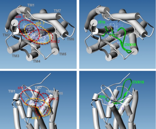
FIGURE 3.
Multiple binding modes for maraviroc into CCR5. All (left) or Glu-283 interacting (right) docked poses of MVC are displayed as thin lines, with CPK color coding of the atomic bonds. The 7TMs of CCR5 are represented by cylinders, as viewed from the extracellular side of the receptor (top) or in the plane of the plasma membrane (bottom). The position of ligands cocrystallized with CXCR4 are indicated in green using ball-and-stick and ribbon representations for the synthetic molecule IT1t and the peptide CVX15, respectively.

