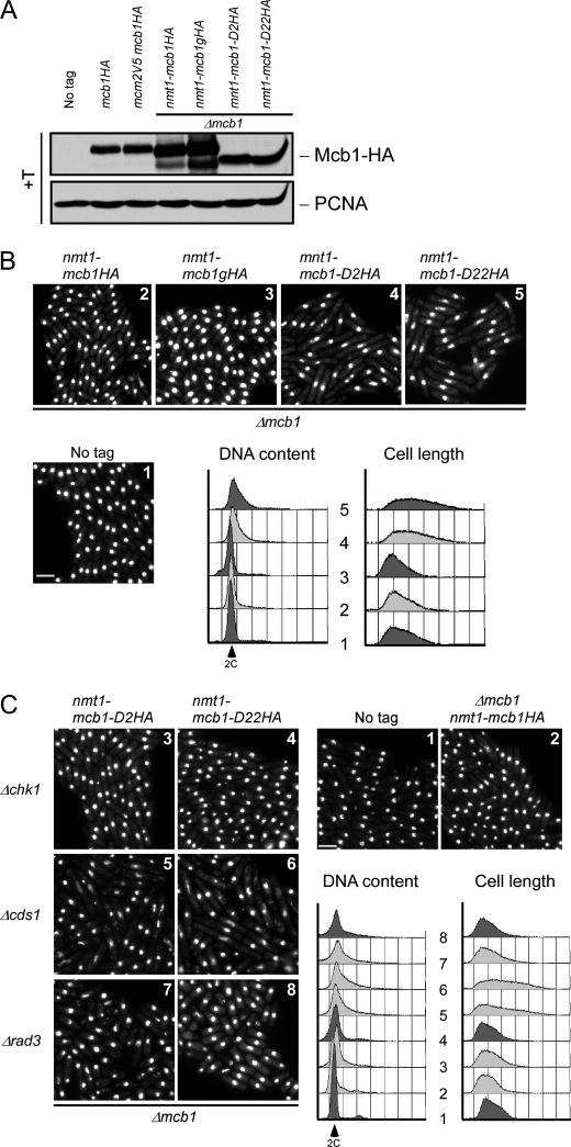
FIGURE 4.
N-terminal deletion mutants (mcb1D2 and mcb1D22) are hypomorphic. A, wild-type (FY11), mcb1HA (FY4041), mcm2V5 mcb1HA (FY4122), Δmcb nmt1-mcb1HA (FY4596), Δmcb nmt1-mcb1gHA (FY5417), Δmcb nmt1-mcb1D2HA (FY5419), and Δmcb nmt1-mcb1D22HA (FY5421) cells were grown asynchronously in medium containing thiamine (+T). An equal number of cells were collected and alkaline lysed. An equal volume of total protein was loaded on an 8% SDS-polyacrylamide gel for separation and immunoblotted for Mcb1HA and proliferating cell nuclear antigen (PCNA) (a loading control). B, photomicrographs 1–5 of DAPI-stained asynchronous wild-type (FY11), Δmcb nmt1-mcb1HA (FY4596), Δmcb nmt1-mcb1gHA (FY5417), Δmcb nmt1-mcb1D2HA (FY5419) and Δmcb nmt1-mcb1D22HA (FY5421) cells. Scale bar, 10 μm. DNA content and cell length were monitored by flow cytometry. C, photomicrographs 1–8 of DAPI-stained wild-type (FY11); Δmcb nmt1-mcb1HA (FY4596); Δmcb nmt1-mcb1D2HA in a Δchk1, Δcds1, or Δrad3 background (FY5499, -5501, or -5505); and Δmcb nmt1-mcb1D22HA in a Δchk1, Δcds1, or Δrad3 background (FY5500, -5503, or -5506) cells. Scale bar, 10 μm. DNA content and cell length were monitored by flow cytometry.

