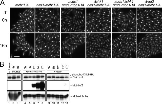
FIGURE 6.
Chk1 is activated in Mcb1-overproducing cells. A, photomicrographs of DAPI-stained mcb1HA (FY4041), nmt1-mcb1HA (FY4594), Δcds1 nmt1-mcb1HA (FY4734), Δchk1 nmt1-mcb1HA (FY4736), Δcds1 Δchk1 nmt1-mcb1HA (FY4739), and Δrad3 nmt1-mcb1HA (FY4740) cells 0 and 16 h after inoculation into −thiamine (−T) medium. Scale bar, 10 μm. B, chk1HA (FY4610) cells were transformed with plasmid expressing Mcb1V5 (pLD18) or empty vector (pSLF972). An equal number of cells were collected at the indicated time points after inoculation into −thiamine medium and alkaline lysed. An equal volume of protein was loaded on an SDS-polyacrylamide gel for separation and immunoblotted for Chk1HA, Mcb1V5, and α-tubulin (a loading control). Lane 1, total lysate of untreated chk1HA cells; lanes 2 and 3, total lysates of chk1HA cells treated with 0.1% methyl methanesulfonate (MMS) for 4 and 1 h (phosphorylated Chk1HA migrates slower), respectively; lanes 4-8, total lysates of pLD18-transformed chk1HA cells from different time points after thiamine removal; and lanes 9-13, total lysates of empty vector (pSLF972)-transformed chk1HA cells from different time points after thiamine removal.

