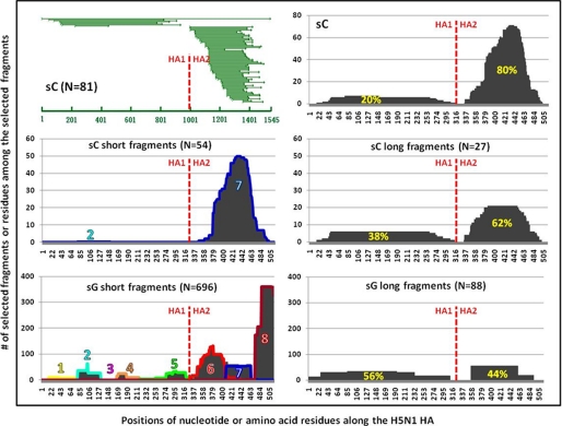
FIGURE 4.
Analysis of antigenic domains based on the positive yeast clones selected by the convalescent plasma samples from the H5N1 (A/Anhui/1/2005 or A/Vietnam/1203/04)-infected humans. Top panel shows the overlapping nucleotide sequences of the positive yeast clones (left panel) and the number of amino acid residues among the selected fragments along their corresponding positions in the HA protein (right panel) (A/Anhui/1/2005). Middle panel shows the antigenic domains in HA protein identified based on the short (left panel) and long (right panel) fragments (A/Anhui/1/2005). Bottom panel shows the antigenic domains identified based on the fragments selected from a phage display library of a different H5N1 strain (A/Vietnam/1203/04) (11). The red vertical line indicates the point where HA1 and HA2 region separate. The percentages highlighted in yellow represent the AUC.

