FIGURE 2.
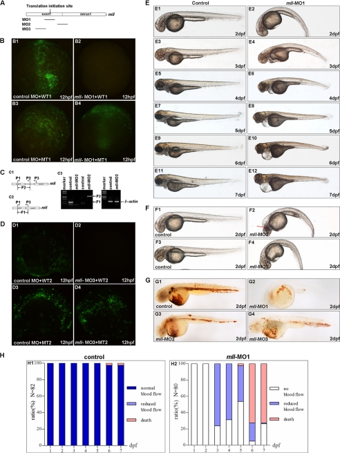
Knockdown of mll by morpholinos (mll-MO1, mll-MO2, and mll-MO3) caused defects in erythropoiesis, blood circulation, and morphogenesis. A, schematic diagram depicts the targets of three morpholinos in zebrafish mll genome. B, validation of mll-MO1. Panel B1, embryos were injected with control MO (8 ng/per embryo) and a wild-type mll (exon1)-GFP fusion protein expression vector (WT1) (5 pg/per embryo) and then examined by fluorescence microscopy. Panel B2, embryos were injected with mll-MO1 (8 ng/per embryo) and a wild-type mll (exon1)-GFP fusion protein expression vector (WT1) (5 pg/per embryo) and then examined by fluorescence microscopy. Panel B3, embryos were injected with control-MO (8 ng/per embryo) and a mismatch-mutated mll-GFP fusion protein expression vector (MT1) (5 pg/per embryo) and then examined by fluorescence microscopy. Panel B4, embryos were injected with mll-MO1 (8 ng/per embryo) and a mismatch mutated mll (exon1)-GFP fusion protein expression vector (MT1) (5 pg/per embryo) and then examined by fluorescence microscopy. C, validation of mll-MO2. Panel C1, schematic diagram depicts the genomic fragment of mll exon1, intron1, and exon2. The positions of PCR primers used to detect fragment 2 (F2) are indicated by arrows. Panel C2, schematic diagram depicts the cDNA fragment of mll exon1 and exon2. The positions of PCR primers to detect fragment 1 (F1) are indicated by arrows. Panel C3, gels show that mll-MO2 (8 ng/per embryo) blocked splicing of exon1 and exon2 of mll efficiently. The fragment 1 resulting from normal splicing was not detected in embryos injected with mll-MO2 (8 ng/per embryo) as indicted in 3rd line (from left to right, left panel), but it was detected in embryos without injection (control) as indicated in 2nd line (from left to right, left panel). Fragment 2 resulting from blocking splicing was detected in embryos injected with mll-MO2 (8 ng/per embryo) as indicated in 5th line (from left to right, left panel), but it was not detected in embryos without injection (control) as indicated in 4th line (from left to right, left panel). The cDNA fragment of β-actin can be detected in both control embryos and mll-MO2 morphants as indicted in 2nd and 3rd lines (from left to right, right panel). D, validation of mll-MO3. Panel D1, embryos were injected with control-MO (8 ng/per embryo) and a wild-type mll(exon1)-GFP fusion protein expression vector (WT2) (5 pg/per embryo) and then examined by fluorescence microscopy. Panel D2, embryos were injected with mll-MO3 (8 ng/per embryo) and a wild-type mll (exon1)-GFP fusion protein expression vector (WT2) (5 pg/per embryo) and then examined by fluorescence microscopy. Panes D3, embryos were injected with control-MO (8 ng/per embryo) and a mismatch-mutated mll-GFP fusion protein expression vector (MT2) (5 pg/per embryo) and then examined by fluorescence microscopy. D4, embryos were injected with mll-MO3 (8 ng/per embryo) and a mismatch mutated mll (exon1)-GFP fusion protein expression vector (MT2) (5 pg/per embryo) and then examined by fluorescence microscopy. E, morphological defects in mll-MO1 morphants. Panel E1, 2-dpf embryos without morpholino injection (control) developed normally. Panel E2, mll-MO1 morphants (2-dpf stage) showed a curved body and heart edema. Panel E3, 3-dpf embryos without morpholino injection (control) developed normally. Panel E4, mll-MO1 morphants (3-dpf stage) showed heart edema and a shortened body. Panel E5, 4-dpf embryos without morpholino injection (control) developed normally. Panel E6, mll-MO1 morphants (4-dpf stage) showed severe heart edema, smaller eyes, and a shortened body. Panels E7, E9, and E11, 5–7-dpf embryos without morpholino injection (control) developed normally. Panels E8, E10 and E12, mll-MO1 morphants (5–7-dpf stage) showed severe heart edema, smaller eyes, a shortened body, and an abnormal head. F, morphological defects in mll-MO2 and mll-MO3 morphants. Panel F1, 2-dpf embryos without morpholino injection (control) developed normally. Panel F2, mll-MO2 morphants (2-dpf stage) showed a curved body and head degenerated. Panel F3, 2-dpf embryos without morpholino injection (control) developed normally. Panel F4, mll-MO3 morphants (2-dpf stage) showed a curved body and heart edema. G, defects of erythropoiesis in mll morphants. Panel G1, erythropoiesis in embryos (2-dpf stage) without morpholino injection was normal as revealed by o-dianisidine staining for hemoglobin. Panel G2, erythropoiesis in embryos (2-dpf stages) injected with mll-MO1 (8 ng/per embryo) was blocked as revealed by o-dianisidine staining for hemoglobin. Panel G3, erythropoiesis in embryos (2-dpf stages) injected with mll-MO2 (8 ng/per embryo) was blocked as revealed by o-dianisidine staining for hemoglobin. Panel G4, the erythropoiesis in embryos (2-dpf stages) injected with mll-MO3 (8 ng/per embryo) was blocked as revealed by o-dianisidine staining for hemoglobin. H, quantitative assays for blood circulation in embryos. Panel H1, different stage embryos without morpholino injection showed normal blood flow. Panel H2, different stage mll-MO1 morphants showed no blood flow or reduced blood flow.

