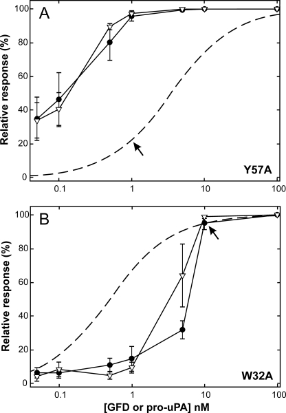
FIGURE 2.
Dose-response curve for GFD and pro-uPAS356A-induced lamellipodia in HEK293 cells transfected with uPARY57A or uPARW32A. Two stably transfected HEK293 cell lines were stimulated with different levels of GFD (▿) or pro-uPAS356A (●), ranging from 0.05 to 100 nm for 24 h, and protrusion indices were subsequently evaluated as outlined under “Material and Methods.” Data shown represent the mean of 6–8 independent experiments for each concentration of added ligand (bars indicate S.E.). To allow direct comparison to ligand occupancy, the protrusion index is depicted as relative scores. The theoretical saturation curves for uPARW32A and uPARY57A (dashed curves) are calculated based on the KD values previously determined by surface plasmon resonance, 0.5 and 3.5 nm, respectively (34). These graphs reveal that HEK293 cells expressing uPARY57A require only ∼20% ligand saturation to accomplish a complete protrusion score (i.e. at 1 nm ligand), whereas cells expressing uPARW32A require 90–95% ligand saturation (i.e. at 10 nm ligand) as indicated by arrows. It is also clear from these experiments that GFD is just as efficient as pro-uPA in stimulating uPAR-dependent protrusions.

