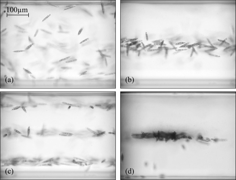
Fig. 3.
Side-view images of acoustic trapping of motile micro-organisms (Euglena gracilis), see Media 1 (4.8MB, MOV) . (a) The acoustic trap is off, the micro-organisms are randomly distributed within the probe volume. (b) Acoustic trap switched on (f = 1.95 MHz), specimens are confined in single nodal plane. (c) Trapping with resonance at f = 5.77 MHz with three nodal planes. (d) Aggregation of specimens due to additional horizontal confinement.

