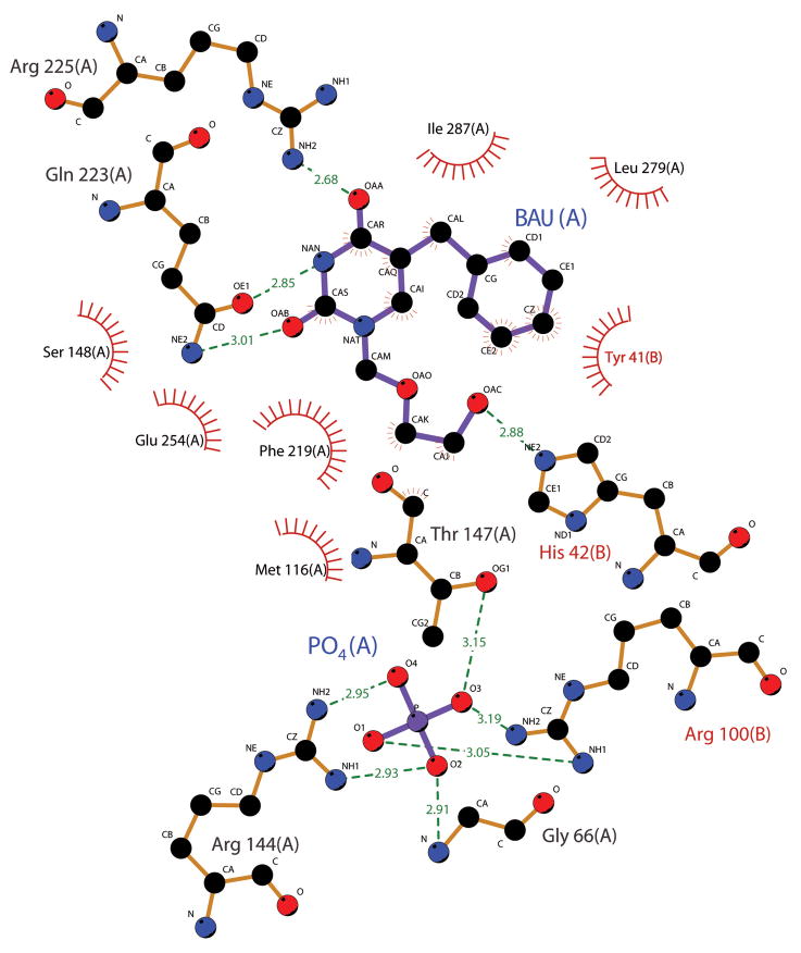
Fig. 3.
Depiction of hUPP2 ligand-interacting residues. Residues forming energetically favorable interactions with either the inhibitor BAU or the phosphate substrate are illustrated using Ligplot (Wallace et al., 1995). All residues shown are strictly conserved in identity and position between hUPP2 and hUPP1.

