
FIGURE 4.
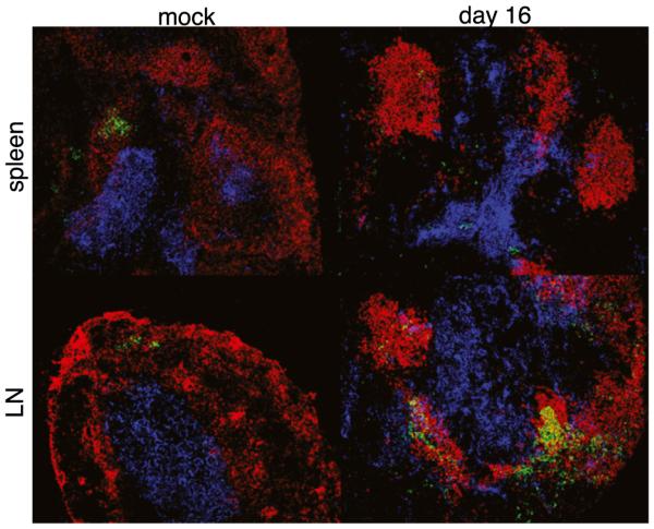
GC formation was observed in the LNs, but not in the spleens, of infected mice. Frozen sections from spleen tissue (top row) and an inguinal lymph node (bottom row) from mock-infected and day 16-infected C57BL/6 mice were stained with Abs specific for B220 (red), Thy1.2 (blue), and PNA (green). Colocalization of the PNA-positive cells with the B and T cells was detected in the LNs, but not in the spleens, of infected mice. Original magnification ×200.