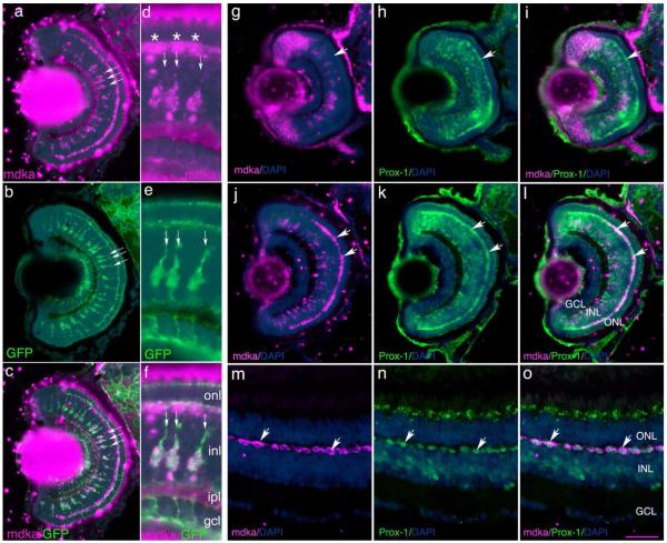
Figure 4. mdka is expressed in Müller glia.
Panels a and d illustrate in situ hybridizations of mdka expression in Tg(gfap:GFP)Mi2001 fish at 120 hpf. Panels b and e illustrate Müller glia immunostained with antibodies against green fluorescent protein. Panel c is the digital overlay of panels a and b; panel f is the digital overlay of panels d and e. In each panel, the three arrows in identify the same three Müller glia. In panel d, the asterisks identify mdka expression in presumptive horizontal cells. Panel g is an in situ hybridizations showing the expression of mdka at 72 hpf. Panel h is the same section as in panel g, but immunostained with antibodies against Prox1. Panel i is the digital overlay of panels g and h. Note that at 72hpf, horizontal cells synthesize Prox1, but do not yet express mdka. Panel j is an in situ hybridization showing the expression of mdka at 120hpf. Panel k is the same section as in panel j, but immunostained with antibodies against Prox1. Panel l is the digital overlay of panels j and k. Note the co-localization of mdka mRNA and Prox1 protein. Panel m is an in situ hybridization showing the expression of mdka in the adult retina. Panel n is the same section as in panel j, but immunostained with antibodies against Prox1. Panel o is the digital overlay of panels d and e. Arrows in j-o identify horizontal cells that express mdka and are immunostained for Prox1. onl, outer nuclear layer; inl, inner nuclear layer; gcl, ganglion cell layer; DAPI, nuclear stain 4,6-diamidino-2-phenylindole dihydrochloride. Scale bar equals 50μm; onl, outer nuclear layer; inl, inner nuclear layer; ipl, inner plexiform layer; gcl, ganglion cell layer; ipl: inner plexiform layer. Scale bar equals 50μm.

