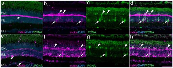
Figure 5. In the light-lesioned retina, mdka and mdkb are expressed by horizontal cells and injury-induced photoreceptor progenitors.
Panel a is an in situ hybridization showing the expression of mdka in a control retina. The white arrow identifies mdka-expressing horizontal cells. Panel b is an in situ hybridization showing mdka expression in a retina following 72 hrs of light exposure. Panel c is the same section as in panel b, immunostained with antibodies against PCNA. Panel d is the digital overlay of panels b and c. Arrowheads and arrows in panels b-d identify double-labeled cells in the ONL and INL, respectively. Panel e illustrates an in situ hybridization showing the expression of mdkb in a control retina. Panel f is an in situ hybridization showing mdkb expression in a retina following 72 hrs of light exposure. The arrowhead and arrow identify mdkb-expressing cells in the inner and outer nuclear layers, respectively. Panel g is the same section as in panel f, immunostained with antibodies against PCNA. Panel h illustrates the digital overlay of panels f and g. In panels b-d and f-h, arrowheads and arrows identify double-labeled cells in the ONL and INL, respectively. ONL, outer nuclear layer; INL, inner nuclear layer; GCL, ganglion cell layer; PCNA, Proliferating Cellular Nuclear Antigen; DAPI: nuclear stain 4,6-diamidino-2-phenylindole, dihydrochloride. Scale bar equals 50μm.

