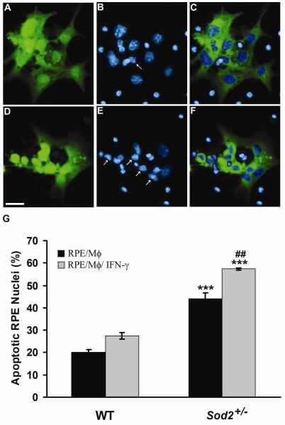
Figure 2. Mononuclear phagocytes induce RPE apoptosis.
Hoechst 33342 staining of WT RPE co-cultured with unstimulated mononuclear phagocytes (A-C) or with IFN-γ-activated mononuclear phagocytes (D-F). WT RPE cells were prelabeled with CellTracker Green CMFDA (A, D). (C) Merged image of prelabeled WT RPE cells (A, green), and nuclei (B, blue) of WT RPE and unstimulated mononuclear phagocytes; (F) Merged image of prelabeled WT RPE cells (D, green), and nuclei (E, blue) of WT RPE and IFN-γ-activated mononuclear phagocytes. WT RPE cells are distinguished from mononuclear phagocytes by merged images (C, F). Arrows in panels B and E indicate condensed WT RPE nuclei. Scale bar, 25 μm. (G) shows percentage of apoptotic nuclei of WT and Sod2+/− RPE cells exposed to unstimulated mononuclear phagocytes (RPE/Mϕ) or IFN-γ-activated mononuclear phagocytes (RPE/Mϕ/ IFN-γ). *** P < 0.001, compared with WT; ##P < 0.01, compared with RPE/Mϕ.

