FIGURE 2.
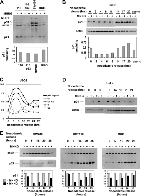
MNNG leads to p21 degradation in a variety of cell lines. A, the indicated cell lines were treated with 10 μm MNNG or dimethyl sulfoxide (DMSO) control for 1 h followed by Western blot analysis of total cell extracts. Bottom panel, quantitation of p21 normalized to β-actin. B, U2OS cells were synchronized by a nocodazole block, released for the indicated times, and then treated with MNNG for 1 h. p21 Western blot is shown. Bottom panel, quantitation of p21 remaining after MNNG treatment and normalized to β-actin. async, asynchronous cells. C, alignment of p21 degradation (p21 degrad) in U2OS with cell cycle analysis; the extent of p21 degradation correlated best with the percentage of cells in G1 plus S phases. D, in HeLa cells, p21 degradation induced by MNNG occurs throughout the cell cycle. E, cell cycle analysis of MNNG-induced p21 degradation in SW480, HCT116, and RKO cells. Bottom panel, quantitation of p21 normalized to β-actin.

