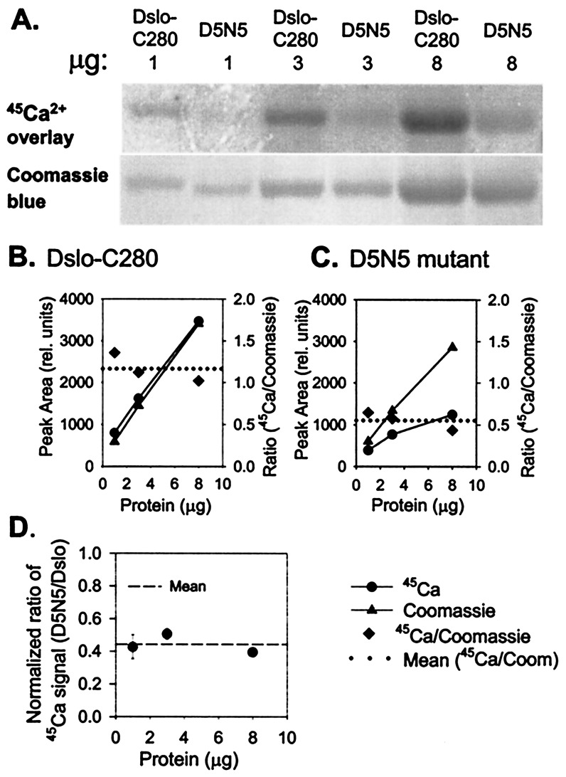
Figure 4.
Comparison of 45Ca2+-binding activity of Dslo-C280 and corresponding D5N5 mutation. (A) Purified Dslo-C280 and D5N5 mutant (1, 3, and 8 μg) were subjected to SDS/PAGE, electroblotted onto PDVF, and assayed by 45Ca2+-overlay method: (Upper) image of exposed film; (Lower) Coomassie blue-stained protein bands. (B and C) Densitometric measurement of protein bands (▴), 45Ca2+ signal (●), and 45Ca2+/Coomassie ratio (♦) for Dslo-C280 (B) and D5N5 mutant (C). (D) Protein-normalized ratio of 45Ca2+-binding activity of D5N5 mutant relative to Dslo-C280.

