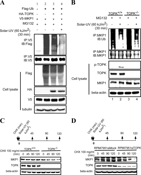
FIGURE 4.
TOPK enhances the stability of MKP1. A, TOPK decreased the ubiquitination of MKP1 in HEK293 cells after treatment with solar UV light (60 kJ/m2). HEK293 cells were transfected with pCMV-FLAG-ubiquitin (Flag-Ub), pcDNA3-HA-TOPK, and pcDNA3-V5-MKP1 as indicated. At 48 h after transfection, cells were stimulated with MG132 (10 μm) for 4 h, followed by exposure to solar UV light (60 kJ/m2) and harvesting 30 min later. The samples were immunoprecipitated (IP) with anti-V5 antibody and detected with anti-FLAG antibody by Western blotting. The transfection efficiency and equal protein loading were verified by Western blotting using the whole cell lysate. IB, immunoblot. B, endogenous ubiquitination of MKP1 in TOPK+/+ MEFs was decreased compared with TOPK−/− MEFs after treatment with solar UV light (60 kJ/m2). TOPK+/+ and TOPK−/− MEFs were pretreated with MG132 (10 μm) for 4 h, followed by stimulation with solar UV light (60 kJ/m2) and harvesting 30 min later. The samples were immunoprecipitated with anti-MKP1 antibody, and ubiquitin was detected with anti-ubiquitin antibody by Western blotting. The phosphorylation of TOPK (Thr-9) and equal protein loading were confirmed by Western blotting using the whole cell lysate. C, the half-life of MKP1 was shorter in TOPK−/− MEFs compared with wild-type cells. TOPK+/+ and TOPK−/− MEFs were stimulated with cycloheximide (CHX; 100 μg/ml) and solar UV light (60 kJ/m2) simultaneously. The protein level of MKP1 was detected by Western blotting. D, the half-life of MKP1 was shorter in RPMI7951 siTOPK melanoma cells compared with RPMI7951 siMock cells. The half-life of MKP1 was assessed in RPMI7951 siMock and RPMI7951 siTOPK melanoma cells as described for C.

