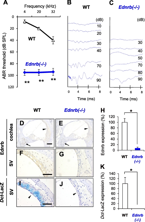
FIGURE 1.
Congenital deafness in Ednrb−/− mice and expression of Ednrb in inner ears. A, hearing levels (means ± S.E. (error bars)) in WT mice (n = 9) and Ednrb−/− mice (n = 9) on P19 measured by ABR. B and C, ABR waveforms of littermate WT mice (WT, B) and Ednrb−/− mice (Ednrb−/−, C) on P19 at 10–90 dB SPL of 12 kHz sound. D–G, immunohistochemical analysis of Ednrb expression in the cochlea (D and E) and the SV (F and G) from Ednrb−/− mice (E and G) and littermate WT mice (D and F) on P19. Arrows and arrowheads in D and E indicate SGNs and the SV, respectively. H, percentage (means ± S.E.) of Ednrb expression in the SV from Ednrb−/− mice (Ednrb−/−, blue bar, n = 3) and littermate WT mice (WT, white bar, n = 3) to that in the SV from WT mice. I and J, LacZ staining of melanocytes in the SV. We employed Dct-LacZ mice, in which the Dct promoter is known as a specific marker of melanocytes (intermediate cells) (31), to establish Dct-LacZ;Ednrb−/− mice newly by crossing Ednrb−/− mice and Dct-LacZ mice (32). K, percentage (means ± S.E.) of LacZ-positive melanocytes in the SV from Ednrb−/− mice (Ednrb−/−, n = 3) and littermate WT mice (WT, white bar, n = 3) to that in the SV from WT mice. LacZ staining showed no positive cells in the SV from Dct-LacZ;Ednrb−/− mice (arrowhead in J and K), whereas Dct-LacZ mice with intact Ednrb showed LacZ-positive melanocytes in the SV (blue signals indicated by arrowhead in I and K). Significant difference (*, p < 0.05; **, p < 0.01) from the control was analyzed by the Mann-Whitney U test. Scale bars: 100 μm (D and E) and 50 μm (F–J).

