FIGURE 2.
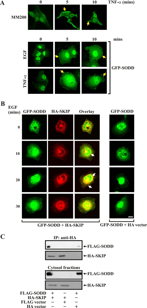
Co-localization of SKIP and SODD. MM200 cells were serum-starved and stimulated with TNF-α for the indicated times, fixed, permeabilized, stained with SODD antibodies, and visualized by confocal microscopy (top panel). COS-7 cells were transfected with GFP-SODD or vector alone for 48 h and then serum-starved and stimulated with either EGF or TNF-α for the indicated times and fixed and visualized by confocal microscopy (bottom panels). Yellow closed arrows indicate plasma membrane localization. B, COS-7 cells were transiently co-transfected with GFP-SODD and HA-SKIP or vector controls, EGF (100 ng/ml)-stimulated, fixed, stained with HA antibodies, and imaged by confocal microscopy. Closed arrows indicate co-localization of GFP-SODD with HA-SKIP. Bar, 20 μm. C, cytosolic fractions of COS-7 cells expressing FLAG-SODD and HA-SKIP or vector controls were prepared as described under “Experimental Procedures,” and HA immunoprecipitates (IP) were analyzed by SDS-PAGE and immunoblotted with FLAG or HA antibodies (top two panels). Cytosol input fractions immunoblotted with either FLAG or HA antibodies are shown in the bottom two panels.

