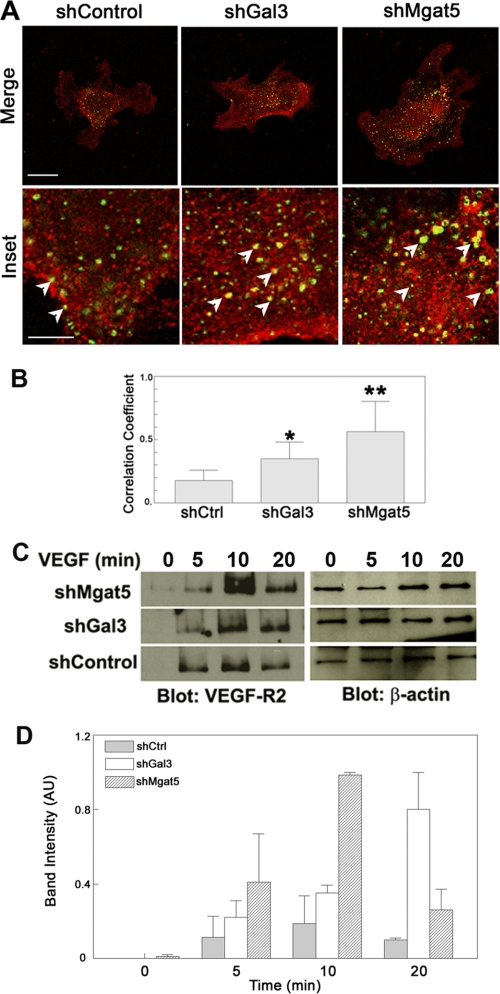
FIGURE 3.
VEGF-R2 internalization is increased in Mgat5 and galectin-3 knockdown cells. To determine whether cells lacking Mgat5 or galectin-3 have increased levels of internalized VEGF-R2, knockdown (shGal3, shMgat5) and shControl endothelial cells were stimulated with VEGF, fixed, permeabilized, probed with anti-VEGF-R2 and anti-EEA1 monoclonal antibodies, and analyzed by confocal microscopy. A, representative images of EEA1- and VEGF-R2-stained HUVE cells. Scale bar = 30 μm; inset scale bar = 10 μm. The arrows indicate areas of VEGF-R2 colocalized with EEA1. The results are representative of two independent experiments. B, Costes correlation coefficient normalized to total EEA1 staining. Data are expressed as mean ± S.E. *, p < 0.01; **, p < 0.05 as compared with shControl cells. The results are representative of two independent experiments (n = at least 5/group). C, to assess VEGF-R2 endocytosis, shControl and knockdown (shMgat5 and shGal3) cells were also biotinylated to label extracellular proteins and treated with VEGF-A to stimulate VEGF-R2 internalization for the indicated time periods. Cell surface bound biotin was then removed by a reducing media wash, and the cells were lysed. The internalized biotinylated proteins, along with whole cell lysates, were separated by SDS-PAGE and analyzed by Western blot analysis for VEGF-R2 and β-actin, respectively. The results are representative of two independent experiments. D, quantification of internalized biotin labeling. Data are expressed mean ± S.E. AU, arbitrary units.

