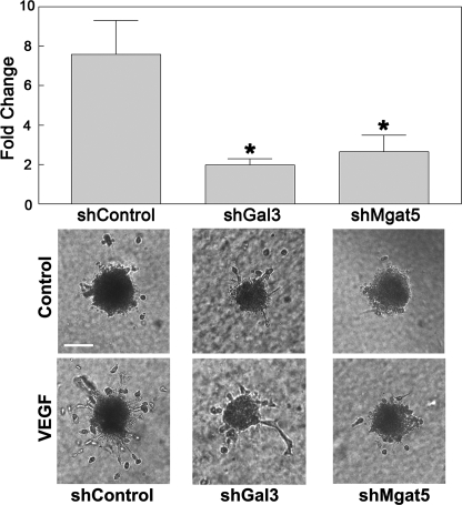
FIGURE 4.
Galectin-3 and Mgat5 knockdown cells display reduced angiogenesis in vitro. To determine the role of galectin-3 and Mgat5 in angiogenesis, an in vitro HUVE cell sprouting assay was used. Endothelial cell spheroids were embedded in a collagen matrix and stimulated with media containing 50 ng/ml VEGF-A. After 24 h, the cumulative length of the sprouts for each spheroid was quantified by ImageJ (top panel). Data are expressed as mean fold change over control-treated cells incubated in media alone ± S.E. *, p < 0.05 as compared with shControl cells. The results are representative of three independent experiments (n = at least 4/group). Also shown are representative images of sprouting spheroids (bottom panel). Scale bar = 100 μm.

