FIGURE 5.
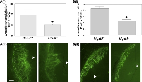
Galectin-3 and Mgat5 knockout mice demonstrate reduced corneal neovascularization. To determine the effect of galectin-3 and Mgat5 on corneal neovascularization, the corneal suture model was used. A, galectin-3 null mice have reduced suture-induced neovascularization. A single suture was placed ∼2 mm above the limbic vessel in the cornea of wild-type (Gal-3+/+) or galectin-3 knockout mice (Gal-3−/−). After 7 days, the mice were perfused intracardially by injection of FITC-BSI and analyzed by fluorescence microscopy. i, the area of fluorescent labeled vessels was measured by ImageJ. Data are expressed as mean ± S.E. (n = 4/group). *, p < 0.05 as compared with Gal-3+/+ mice. The results are representative of two independent experiments. ii, representative fluorescence images of corneas. The arrowheads indicate sutures. Scale bar = 100 μm. B, Mgat5 knockout mice demonstrate reduced corneal neovascularization. i, corneal neovascularization in wild-type (Mgat5+/+) and Mgat knock-out mice (Mgat5−/−) was evaluated using the corneal suture model. Data are expressed as mean ± S.E. (at least n = 4/group). *, p < 0.05 as compared with Mgat5+/+ mice. The results are representative of two independent experiments. ii, representative fluorescence images of corneas. The arrowheads indicate sutures. Scale bar = 200 μm. BS1, Bandeira simplicifolia.

