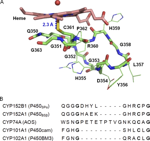
FIGURE 5.
The proximal side of the heme of P450SPα. A, the main chain composed of the loop at the proximal side of the heme is represented as a green stick model. Heme is shown as a pink stick model. The water molecule on the heme is represented as a red sphere. The distances between the heme iron and nitrogen atoms of the main chain from the sulfur atom of Cys361 are shown in blue. B, comparison of the amino acid sequence of P450SPα with other P450s. Conserved amino acid residues are depicted in bold type.

