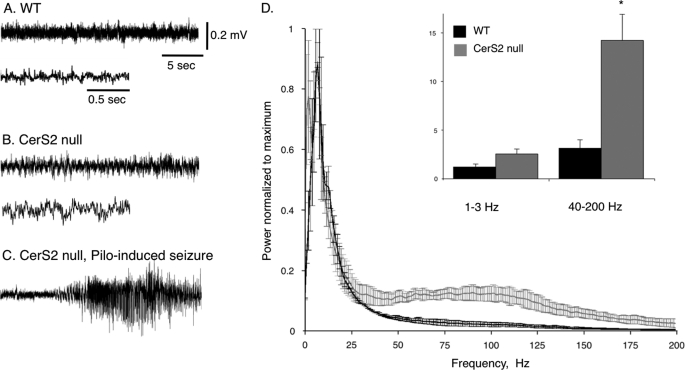
FIGURE 9.
EEG analysis of motor dysfunction. EEG traces were recorded using epidural electrodes in WT (A) and CerS2-null (B) mice. Recording in the CerS2-null mice was performed during myoclonic jerk movements. The lower trace in B shows the same recording but with a faster time scale. C, a typical epileptic seizure was recorded 2 min after injection of the muscarinic agonist pilocarpine (Pilo) (310 mg/kg), confirming that the implanted electrodes are able to record cortical seizures (36). D, power spectrum density. The integral of activity for each mouse was calculated (n = 5) for slow δ (1–3 Hz) and fast γ (40–200) frequency ranges. Note the significant increase in fast γ activity in the cortex of CerS2-null mice. *, p < 0.01.

