FIGURE 3.
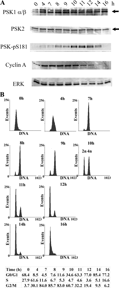
PSKs are activated catalytically and phosphorylated during mitosis. Growing HeLa cells were synchronized using a double thymidine block and then released from G1/S into the cell cycle. A, at the times shown after thymidine removal cell cultures were lysed and immunoblotted with antibodies to detect PSK1-α/β, PSK2, PSK-Ser(P)-181, cyclin A, or ERK. Arrows indicate PSK1-α/β (lower band) or PSK2 (upper band) (as confirmed using siRNA knockdown elsewhere in the manuscript) (“Results” and Fig. 7C). B, alternatively, after thymidine removal, cell cultures were fixed in 70% ethanol and stained with propidium iodide to determine cell cycle profiles using FACS. The percentages of cells in G0/G1, S and G2/M are shown.

