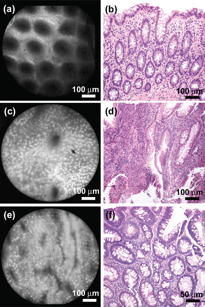
Figure 2.
HRME imaging in the colon. (a) HRME image of the normal colon in vivo, with corresponding histopathology from the same site (b). Note the appearance of small, uniformly spaced circular crypts, with small, basally oriented nuclei. (c) HRME image and (d) corresponding histopathology of an inflammatory polyp, presenting a dense population of inflammatory cells and few irregularly shaped glands. (e) HRME image of a tubular adenoma, revealing highly irregular and heterogeneously oriented glands, with elongated, crowded cells, and enlarged nuclei.

