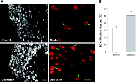
Fig. 2.
Localization of P2X3 in lumbar DRG neurons 24 h following femoral artery occlusion. Fluorescence immunohistochemistry was employed to examine expression of P2X3 in DRG neurons of control limb and occluded limb. A: representative photomicrographs show P2X3 staining at a lower power (left) and a higher power (right). Arrows indicate P2X3-positive cells. Scale bar = 50 μM. B: histograms show that percentage of P2X3-positive neurons is greater in DRG neurons of femoral artery occlusion (n = 6) than that in control (n = 6). *P < 0.05, compared with control.

