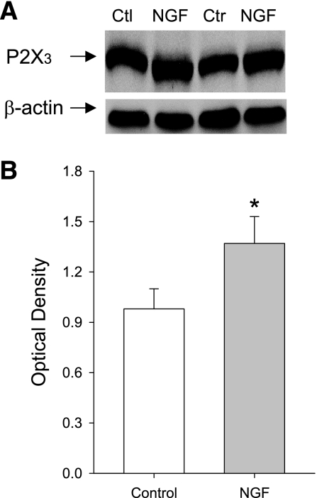
Fig. 5.
Effects of nerve growth factor (NGF) infusion on expression of P2X3 proteins in DRG neurons. Exogenous NGF was continually infused at a rate of 0.25 μg/h into the hindlimb of healthy rats with a microosmotic pump for 72 h. Saline was infused into the contralateral limb as control. Once deliveries of NGF and saline were complete, the levels of P2X3 proteins were examined in bilateral DRGs (L4–6) using western blot analysis. A: representative bands of P2X3 expression. Bands of β-actin are used as control for an equal protein loading. Note that the repetitive control (Ctl, Ctr) and NGF expressions are shown. B: average data. Optical density is expressed in arbitrary units normalized against a control sample. Data in histograms represent means ± SE; n = 6 animals per group. *P < 0.05, compared with control.

