Fig. 8.
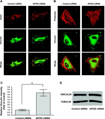
Focal adhesions are increased when APOD expression is attenuated. HDFNs were transfected with control siRNA or APOD siRNA to knockdown APOD expression. Forty-eight hours posttransfection, cells were plated on 10 μg/ml collagen I and allowed to attach for 30 min before fixing for immunostaining. A: cells were immunostained to visualize focal adhesions using zyxin and vinculin antibodies. B: cells were stained with fluorescently labeled phalloidin to visualize stress fibers, and vinculin to mark focal contacts. Pictures were captured at ×400 magnification. C: focal adhesions were quantified using vinculin staining intensity. *P < 0.0001. D: Western blot to detect vinculin expression in siRNA control and APOD-treated cells. Tubulin was used as a control to show equivalent amounts of total protein.

