Fig. 1.
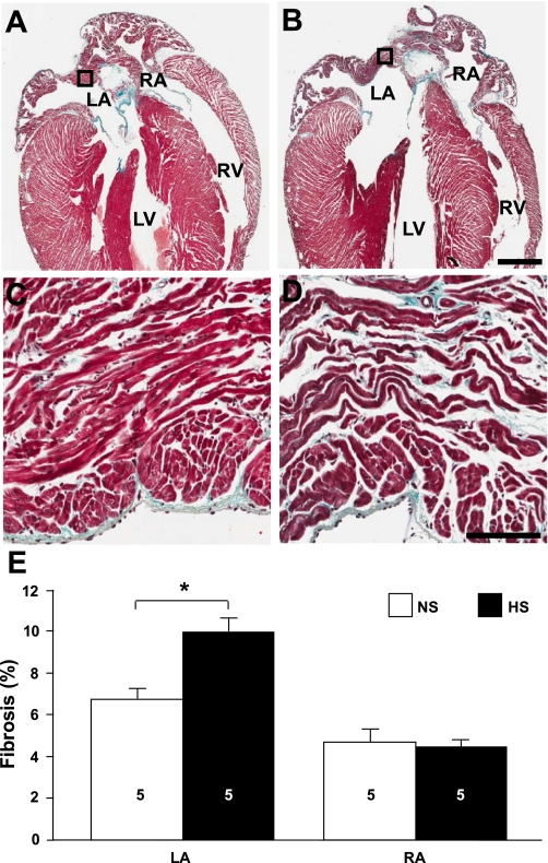
Increased left atrial (LA) fibrosis in high-salt diet-fed (HS) animals. A and B: trichrome-stained sections of hearts from normal-salt diet-fed (NS; A) and HS (B) animals. Bar = 1 mm. RA, right atrium; LV, left ventricle; RV, right ventricle. C and D: high-magnification images of the boxed regions indicated in A and B, respectively. Bar = 0.1 mm. E: average fibrosis values for LA and RA free walls and appendages (LAA and RAA, respectively). Numbers in bars indicate numbers of animals in each group. *P < 0.05.

