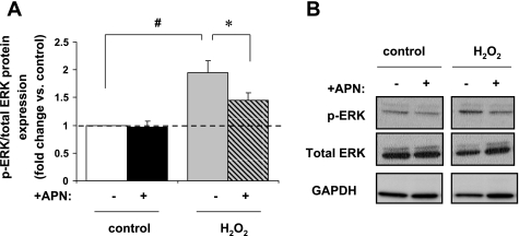
Fig. 3.
In vitro protein expression of p-ERK MAPK. A: H2O2 (10 μM/15 min) increased p-ERK protein expression by 2.2 ± 0.3-fold (#P < 0.01 vs. control). This increase was diminished to 1.5-fold with APN pretreatment (*P < 0.05 vs. H2O2 alone; n = 7). Results are expressed relative to control. B: representative Western blot of p-ERK expression. Protein expression of p-ERK is normalized to total ERK.

