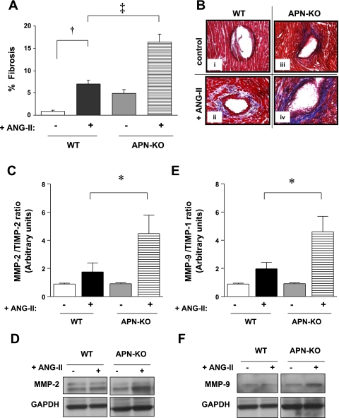
Fig. 7.
In vivo cardiac fibrosis. A: ANG II infusion (3.2 mg·kg−1·day−1 for 14 days) increased fibrosis in WT LV by 7.9-fold (†P < 0.001 vs. WT saline; 7.0 ± 1% vs. 0.9 ± 0.2%). ANG II infusion further enhanced fibrosis in APN-KO (16.4 ± 1.8%) by 2.4-fold (‡P < 0.001 vs. WT ANG II). Data reflect measurements of three sections each for 3 WT saline, 3 APN-KO saline, 6 WT ANG II, and 5 APN-KO ANG II mice. B: representative light microscopy images (×400 magnification); ANG II infusion increased fibrosis in WT ventricles (ii, indicated by aniline blue stain) compared with control (i). LV from APN-KO mice exhibit enhanced fibrosis in response to ANG II infusion (iv) vs. ANG II-infused WT mice (ii). C: ANG II infusion significantly increased the MMP-2-to-TIMP-2 ratio in the LV of APN-KO (*P < 0.05 vs. WT ANG II). D: representative Western blot of MMP-2 expression. E: ANG II infusion significantly increased the MMP-9-to-TIMP-1 ratio in the LV of APN-KO. F: representative Western blot of MMP-9 expression. (*P < 0.05 vs. WT ANG II; n = 3–6).

