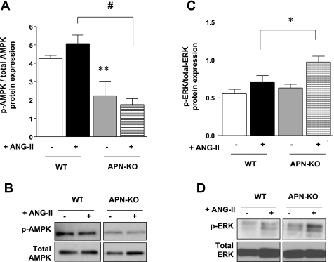
Fig. 8.
In vivo protein expression of p-AMPK. A: ventricular p-AMPK expression was significantly decreased in APN-KO ANG II mice by 105 ± 4% (#P < 0.001 vs. WT ANG II), although there was no difference in p-AMPK expression between saline and ANG II-infused hearts of both WT and APN-KO mice. B: representative Western blot of p-AMPK and total AMPK expression. C: ventricular p-ERK expression was increased in APN-KO ANG II mice by 38 ± 5%. D: representative Western blot of p-ERK and total ERK expression. (*P < 0.05 vs. WT ANG II; n = 3–6).

