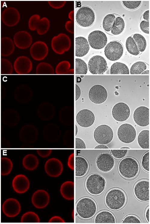
Figure 2. Mitocondrial functionality of developing sea urchin embryos.
(A,C and E) Active mitochondria revealed by the mitochondrial-specific fluorescent dye Mitotracker after 50 min post-fertilization. (B,D and F) Corresponding bright field images. (A) Control embryos. (C) Embryos incubated with 5 µg/ml DD; (E) embryos incubated with 5 µg/ml DD in the presence of the peroxynitrite scavenger MnTBAP (10 µM).

