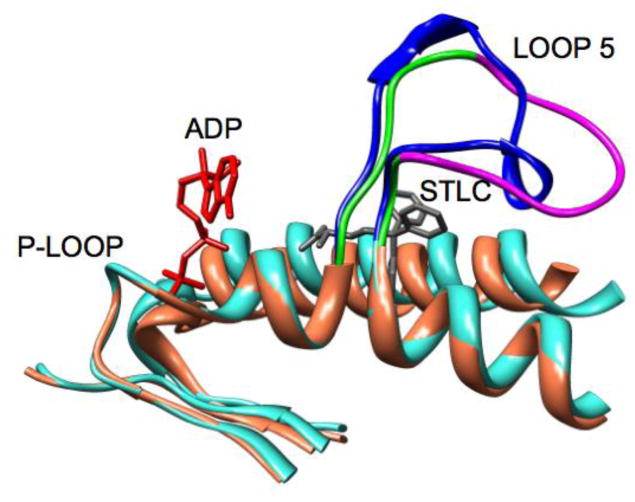
Figure 2.
An overlay of ribbon diagrams of the structures of Eg5•ADP [10] and Eg5•ADP with bound STLC [13]. Eg5•ADP is colored coral except for L5, which is colored green and magenta. The magenta segment is the portion of L5 that is removed in the Eg5-ΔL5 construct. Eg5•ADP with bound STLC is colored turquoise, except for L5, which is blue. STLC is gray. ADP is red.

