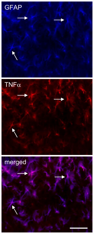
Figure 4.
Determination of cellular localization of TNFα in the spinal dorsal horn in rats treated with ddC for 2 weeks. Immunostaining of GFAP and TNF3 was carried out. There was an almost complete colocalization between GFAP and TNFα imaging, but TNFα did not colocalize with NeuN (data not shown), which suggested that TNFα was located in the astrocytes, but not neurons. Arrow shows the colocalization. Scale bar, 100μm.

