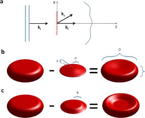
Fig. 1.

(a) Plane wave illumination of a blood smear; ki and ks are the incident and scattered wave vectors. (b) The discocyte modeled by subtracting two Gaussian surfaces from the top and bottom of a cylinder. c) A flat top Gaussian describes deflated cells as described in Section. 4.
