
Fig. 2.
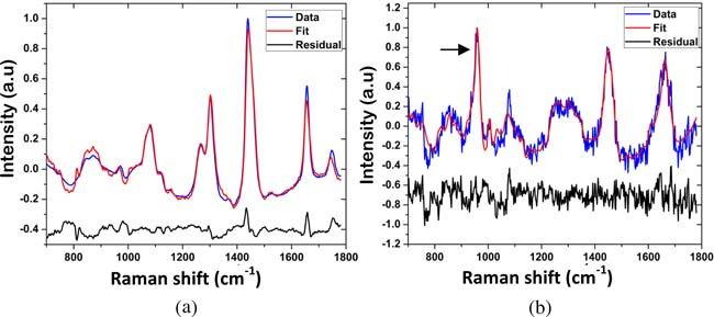
Typical Raman spectra of (a) normal breast tissue and (b) breast lesion (fibrocystic change) with type II microcalcifications, with model fits and residuals. A prominent band at 960 cm-1 due to hydroxyapatite (arrow) is present in the Raman spectrum of the lesion with microcalcifications.