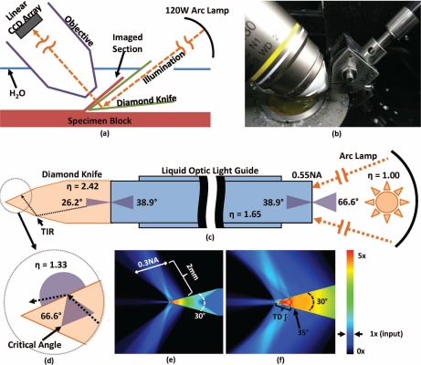
Fig. 1.
Knife-edge scanning microscopy. Serial sections are concurrently cut and imaged under water (a–b). Imaging is performed in transmission mode by sending illumination through a diamond cutting tool. Image capture is synchronized with stage position and performed using a line-scan camera. Illumination is provided using a mercury vapor short arc lamp to send light to the knife through a liquid-optic light guide (0.55NA, η = 1.65). Geometric angles of the refracted light are shown for each interface (c). Total internal reflection (TIR) within the diamond knife at the diamond/water interface results in light scattering and emission through the knife tip when the incident angle is less than the critical angle (33.3°) for a diamond knife (η = 2.42) immersed in water (η = 1.33) (d). The density of illumination around the knife is estimated using a first-order Monte Carlo simulation (e–f). The amount of scattering is determined by the NA of the light guide. Light intensity is shown as a factor of input intensity using the associated color map. Imaging is performed using time-delayed integation (TDI) as the tissue moves along the 35° bevel at the knife tip. Using TDI increases the SNR and increases the amount of scattered illumination used to construct the final image.

