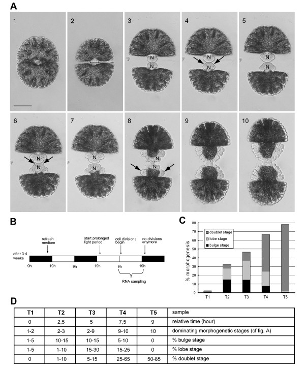
Figure 1.
Morphogenesis of Micrasterias denticulata and distribution of morphogenetic stages in the synchronized sample series. (A) Morphogenesis of M. denticulata. (1) Vegetative cell. (2) During mitosis, a septum originating from the cell wall girdle grows inward centripetally, taking 15-20 min. (3) Bulge stage; the septum bulges uniformly. (4) Development of the first pair of indentations (arrows), ~75 min after septum completion. (5) Three-lobed stage. (6) Development of the second pair of indentations (arrows). (7) Five-lobed stage. (8) Doubling of the lateral lobes (arrows). (9) Formation of further indentations and lobe tips, followed by the doublet stage. N, Nucleus. Note the migration of the nucleus during cell growth. Scale bar = 100 μm. (B) Scheme of the synchronization protocol. After 3-4 weeks, a stationary culture is obtained and the growth medium is refreshed, concomitantly with the reduction in cell density, shortly before the beginning of the light period of that day. The majority of the cells divide in the second dark period afterward. This dark period is replaced by a light period and sampled. Black, dark period; white, light period. (C), Distribution of morphogenetic stages in the RNA samples for cDNA-AFLP, replication 1. (D) Table representing the characteristics of the samples used for cDNA-AFLP (replications 1 and 2) and real-time qPCR.

