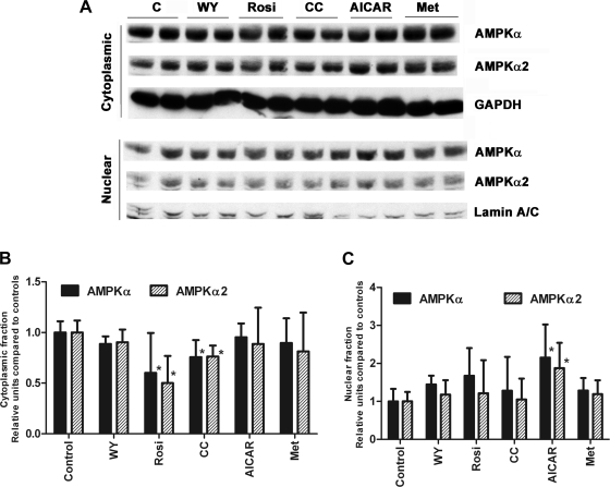
Fig. 4.
A: H4IIEC3 cells were subjected to the treatments as described for 24 h before harvesting and cellular fractionation. Nuclear and cytoplasmic fractions (10 μg) were subjected to Western blotting for AMPK-α and -α2. B: cytoplasmic levels of total AMPK-α and -α2. Protein levels were normalized to GAPDH. *P < 0.05 compared with control. C: nuclear levels of total AMPK-α and α-2. Protein levels were normalized to the levels of lamin. *P < 0.05 compared with control. N = 3.

