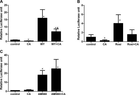
Fig. 6.
H4IIEC3 cells were transfected with the reporter plasmids and PPAR-α (A) or -γ (B), and the AMPK-α1312 expression plasmid as noted by CA (constitutively active). The cells were treated with WY, rosiglitazone, and AM580 at the same concentrations noted in Figs. 1–3 for 24 h. A: PPAR-α, *P < 0.05 compared with control; $P < 0.05 compared with WY-stimulated cells. B: PPAR-γ, *P < 0.05 compared with control. C: RAR. *P < 0.05 compared with control.

