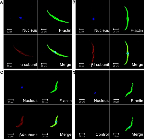
Fig. 3.
Immunocytochemical detection of BK channel α-, β1-, and β4-subunits in freshly isolated human DSM cells using specific antibodies. Red staining (bottom left) indicates detection of the α-subunit (A), β1-subunit (B), and β4-subunit (C). Cell's nuclei are shown in blue (top left); F-actin is shown in green (top right). The merged images are also shown (bottom right) and illustrate overlap of nucleus, F-actin, and the expected BK channel subunit. In control experiments (D), the primary antibody was omitted, and cells were incubated only with the secondary antibody (Control). Images at ×63 were acquired with a Zeiss LSM 510 META confocal microscope. The slides for each group were imaged with the same laser power, gain settings, and pinhole for the controls and antibody treatment. Results were verified in 9 DSM cells freshly isolated from 3 patients.

